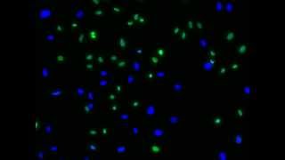
Media Summary: MIT 7.016 Introductory Biology, Fall 2018 Instructor: Adam Martin View the complete course: Discover how we visualized the Cellaca MX and Cellaca PLX high-throughput Many factors can influence the results of your life

Readyprobes Simplifying Cell Imaging - Detailed Analysis & Overview

MIT 7.016 Introductory Biology, Fall 2018 Instructor: Adam Martin View the complete course: Discover how we visualized the Cellaca MX and Cellaca PLX high-throughput Many factors can influence the results of your life

Photo Gallery

ReadyProbes® - Simplifying Cell Imaging
5.5 Ethical considerations–Fixed cell imaging: 5 steps for publication-quality images
29. Cell Imaging Techniques
1.5 Autofluorescence–Fixed cell imaging: 5 steps for publication-quality images
Five steps for publication-quality fixed-cell imaging the first time Complete Webinar
3.4 Controls–Fixed cell imaging: 5 steps for publication-quality images
5.1 Imaging platforms-hardware–Fixed cell imaging: 5 steps for publication-quality images
1.4 Blocking–Fixed cell imaging: 5 steps for publication-quality images
4.2 Photobleaching and antifades–Fixed cell imaging: 5 steps for publication-quality images
3.3 Amplification techniques–Fixed cell imaging: 5 steps for publication-quality images
1.1 Culture conditions–Fixed cell imaging: 5 steps for publication-quality images
Time course of cell death visualized using ReadyProbes™ reagents.
View Detailed Profile
ReadyProbes® - Simplifying Cell Imaging

ReadyProbes® - Simplifying Cell Imaging

ReadyProbes

5.5 Ethical considerations–Fixed cell imaging: 5 steps for publication-quality images

5.5 Ethical considerations–Fixed cell imaging: 5 steps for publication-quality images

https://www.thermofisher.com/us/en/home/life-science/cell-analysis/

29. Cell Imaging Techniques

29. Cell Imaging Techniques

MIT 7.016 Introductory Biology, Fall 2018 Instructor: Adam Martin View the complete course: https://ocw.mit.edu/7-016F18 ...

1.5 Autofluorescence–Fixed cell imaging: 5 steps for publication-quality images

1.5 Autofluorescence–Fixed cell imaging: 5 steps for publication-quality images

https://www.thermofisher.com/us/en/home/life-science/cell-analysis/

Five steps for publication-quality fixed-cell imaging the first time Complete Webinar

Five steps for publication-quality fixed-cell imaging the first time Complete Webinar

https://www.thermofisher.com/us/en/home/life-science/cell-analysis/

3.4 Controls–Fixed cell imaging: 5 steps for publication-quality images

3.4 Controls–Fixed cell imaging: 5 steps for publication-quality images

https://www.thermofisher.com/us/en/home/life-science/cell-analysis/

5.1 Imaging platforms-hardware–Fixed cell imaging: 5 steps for publication-quality images

5.1 Imaging platforms-hardware–Fixed cell imaging: 5 steps for publication-quality images

https://www.thermofisher.com/us/en/home/life-science/cell-analysis/

1.4 Blocking–Fixed cell imaging: 5 steps for publication-quality images

1.4 Blocking–Fixed cell imaging: 5 steps for publication-quality images

http://www.thermofisher.com/us/en/home/life-science/cell-analysis/

4.2 Photobleaching and antifades–Fixed cell imaging: 5 steps for publication-quality images

4.2 Photobleaching and antifades–Fixed cell imaging: 5 steps for publication-quality images

https://www.thermofisher.com/us/en/home/life-science/cell-analysis/

3.3 Amplification techniques–Fixed cell imaging: 5 steps for publication-quality images

3.3 Amplification techniques–Fixed cell imaging: 5 steps for publication-quality images

https://www.thermofisher.com/us/en/home/life-science/cell-analysis/

1.1 Culture conditions–Fixed cell imaging: 5 steps for publication-quality images

1.1 Culture conditions–Fixed cell imaging: 5 steps for publication-quality images

http://www.thermofisher.com/us/en/home/life-science/cell-analysis/

Time course of cell death visualized using ReadyProbes™ reagents.

Time course of cell death visualized using ReadyProbes™ reagents.

Time course of

Revvity Cellaca MX & PLX | 3D Animation for Cell Counting and Image Cytometry | REBUS Agency

Revvity Cellaca MX & PLX | 3D Animation for Cell Counting and Image Cytometry | REBUS Agency

Discover how we visualized the Cellaca MX and Cellaca PLX high-throughput

Viability determination of HeLa cells using ReadyProbes® Cell Viability Imaging Kit (Blue/Green)

Viability determination of HeLa cells using ReadyProbes® Cell Viability Imaging Kit (Blue/Green)

Learn more at http://www.lifetechnologies.com/

3.1 Secondary antibody choice–Fixed cell imaging: 5 steps for publication-quality images

3.1 Secondary antibody choice–Fixed cell imaging: 5 steps for publication-quality images

https://www.thermofisher.com/us/en/home/life-science/cell-analysis/

Best practices: 5 steps to live-cell imaging

Best practices: 5 steps to live-cell imaging

https://www.thermofisher.com/us/en/home/life-science/cell-analysis/

5.2 Imaging platforms-software–Fixed cell imaging: 5 steps for publication-quality images

5.2 Imaging platforms-software–Fixed cell imaging: 5 steps for publication-quality images

https://www.thermofisher.com/us/en/home/life-science/cell-analysis/

2.1 Primary antibody choice–Fixed cell imaging: 5 steps for publication-quality images

2.1 Primary antibody choice–Fixed cell imaging: 5 steps for publication-quality images

https://www.thermofisher.com/us/en/home/life-science/cell-analysis/

Preparing Adherent Cells for Live Cell Imaging

Preparing Adherent Cells for Live Cell Imaging

Many factors can influence the results of your life

Viability determination of HeLa cells using ReadyProbes® Cell Viability Imaging Kit (Blue/Red)

Viability determination of HeLa cells using ReadyProbes® Cell Viability Imaging Kit (Blue/Red)

Learn more at http://www.lifetechnologies.com/